In pediatric traumatology, there are routines that seem almost automatic.
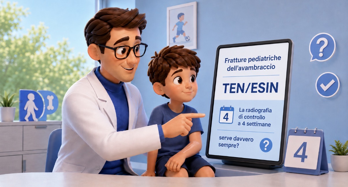
One of these is the radiographic check-up at approximately 4 weeks after osteosynthesis with TEN/ESIN for radius and ulna fractures.
A child who has undergone surgery, with a smooth recovery, improving pain, wounds healing well, and good hand movement. The outpatient check-up arrives, and often the question is already implicit:
"Should we do an X-ray?"
A recently published study in the Journal of Pediatric Orthopaedics sought to understand how much this habit truly changes patient management [1].
The authors retrospectively analyzed children treated with ESIN for forearm fractures, evaluating a very concrete aspect: in clinically asymptomatic patients, did routine 4-week radiography alter treatment?
The study's answer was essentially no [1].
The point is not "to do fewer check-ups"
It's important to clarify this immediately, because the risk of oversimplification always exists.
The study does not say that X-rays are useless.
It says something different: in children who are clinically well, some X-rays performed only "by protocol" may not add information capable of changing therapy.
These are two very different concepts.
In the outpatient clinic, it's common to see children who arrive for their check-up already almost recovered from a clinical point of view:
- minimal pain,
- good finger mobility,
- no new trauma,
- parents at ease,
- gradual return to daily activities.
In these cases, it's natural to wonder whether follow-up should be guided primarily by clinical signs or by the calendar.
Increasingly "risk-adapted" medicine
In recent years, even in pediatric orthopedics, we are witnessing a fairly evident cultural shift.
For a long time, follow-up was organized with standardized protocols identical for everyone:
- check-up at X days,
- X-ray at Y weeks,
- another X-ray at Z months.
Today, however, there is growing attention towards more personalized pathways based on real risk.
The study proposes precisely this approach: not to eliminate check-ups, but to adapt them to the characteristics of the patient and the fracture [1].
For example, probably not all cases have the same risk profile:
- very stable vs. borderline synthesis,
- cooperative child vs. poor compliance,
- isolated trauma vs. new fall,
- perfectly calm clinical picture vs. persistent pain.
And this is where clinical evaluation remains fundamental.
A very common situation
Let's consider a typical scenario.
A 10-year-old child, diaphyseal radius and ulna fracture treated with TEN.
Regular postoperative check-up. After a few weeks:
- almost no pain,
- well-perfused hand,
- normal finger mobility,
- no intermediate trauma,
- linear course.
In such a situation, does routine radiography really change anything in management? This is exactly the question this study tries to address.
Of course, there are also different situations where radiographic control remains absolutely important:
- increasing pain,
- suspected loss of reduction,
- new fall,
- neurological symptoms,
- doubts about initial stability,
- unconvincing clinical course.
Clinical assessment, once again, guides the reasoning.
Fewer X-rays does not mean less attention
This is perhaps the most important message to convey to families as well.
Sometimes "doing everything" is perceived as synonymous with better care. But in modern medicine, the issue is not the quantity of tests: it's understanding which tests truly change decisions.
Reducing unnecessary X-rays also means:
- less radiation exposure,
- fewer hospital visits,
- fewer school and work absences,
- lower indirect costs,
- less medicalization of the recovery process.
Obviously, caution is needed. The study is retrospective and does not allow for absolute conclusions [1]. Furthermore, in real practice, there are variables that are difficult to standardize:
- surgeon's experience,
- type of fracture,
- quality of synthesis,
- reliability of follow-up,
- child's athletic level.
However, the underlying message is interesting: perhaps some historical habits deserve critical re-evaluation.
Not to "do less."
But to do what is truly necessary.
References
[1] Mojica Crespo RM, Memmel C, Hundhausen L, et al. Are Routine 4-Week Radiographs After ESIN Osteosynthesis in Pediatric Forearm Fractures Necessary?: A Retrospective Analysis and Proposal of a Risk-Adapted Follow-up Protocol. Journal of Pediatric Orthopaedics. 2026. doi:10.1097/BPO.0000000000003321
PubMed: https://pubmed.ncbi.nlm.nih.gov/42102347/
Disclaimer: content for general informational purposes only. It does not replace a medical evaluation.
