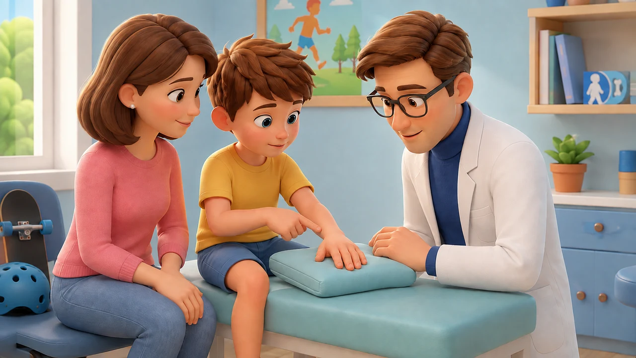
A normal X-ray after an injury is generally reassuring: it shows no obvious fractures, dislocations, or other bone alterations recognizable in the images taken at that time.
In children, however, some injuries may be poorly visible on initial X-rays, and many causes of pain involve structures that X-rays only partially study. If pain persists, the subsequent decision depends mainly on the location, clinical examination, and evolution of symptoms.
Occult Fractures
A fracture can be present even when the first X-ray does not show an obvious fracture line.
In hand or wrist trauma, for example, ACR guidelines consider a repeat X-ray after a period, an MRI, or a CT scan as appropriate options when clinical suspicion remains high. [1]
At the elbow, indirect findings such as a fat pad sign can be associated with an occult fracture. A meta-analysis confirmed that the risk is not negligible in children with this finding, but the image must always be interpreted together with the physical examination. [2]
The Pediatric Skeleton Is Different
Growth plates and structures that are still predominantly cartilaginous make some injuries less obvious compared to the adult skeleton. This is particularly true in young children. [3]
The interpretation of the X-ray must therefore take into account the child's age and the ossification centers present in the studied area.
Pain Without Obvious Fracture
After an injury, pain can also originate from joints, ligaments, tendons, muscles, cartilage, or bone edema.
In the knee, for example, MRI is useful when it is necessary to study bone marrow, menisci, ligaments, and other internal structures that X-rays do not adequately show. [4]
This type of in-depth investigation is not necessary after every negative X-ray. It is chosen when the examination or the course of symptoms maintains a specific doubt.
Evolution in the Following Days
A child who progressively uses the limb better, has less pain, and resumes activities follows a different path from one who continues not to bear weight, maintains very localized pain, or worsens.
Clinical re-evaluation often allows deciding whether more time is sufficient or if the initial hypothesis needs to be revised.
Repeating the X-ray
In some fractures, the signs become more evident after a few days. In suspected hand or wrist trauma with initial negative X-rays, the ACR also considers a repeat X-ray after 10–14 days to be appropriate. [1]
In other areas, repeating the same examination may add little, and it is better to choose a different method. Therefore, there is no standard interval valid for every injury.
MRI, CT, and Ultrasound
Magnetic resonance imaging (MRI) effectively studies bone marrow, cartilage, joints, and soft tissues without ionizing radiation.
CT offers very detailed bone definition and is useful in selected situations, but it involves radiation exposure.
Ultrasound can effectively answer some questions and in certain areas, but it does not generally replace X-rays or MRI.
The choice of examination depends on the structure suspected to be injured and what would change in management.
When to Re-evaluate the Child
Very localized pain that does not improve, persistent difficulty using an arm or bearing weight on a leg, significant swelling, or a course different from what was expected are reasonable reasons for a new evaluation.
Often, re-evaluation confirms that the evolution is normal and no further tests are needed. In other cases, it allows recognizing an injury that was initially not obvious or directing imaging towards a structure other than bone.
References
- American College of Radiology. ACR Appropriateness Criteria® Acute Hand and Wrist Trauma. ACR
- Kappelhof M, et al. Occult Fractures in Children with a Radiographic Fat Pad Sign of the Elbow: A Meta-Analysis of 10 Published Studies. JBJS Reviews. 2022. PubMed
- DeFroda SF, et al. Radiographic Evaluation of Common Pediatric Elbow Injuries. Orthopedic Reviews. PMC
- American College of Radiology. ACR Appropriateness Criteria® Acute Trauma to the Knee. PubMed
Disclaimer: This content is for informational purposes only and does not replace an individual clinical evaluation.