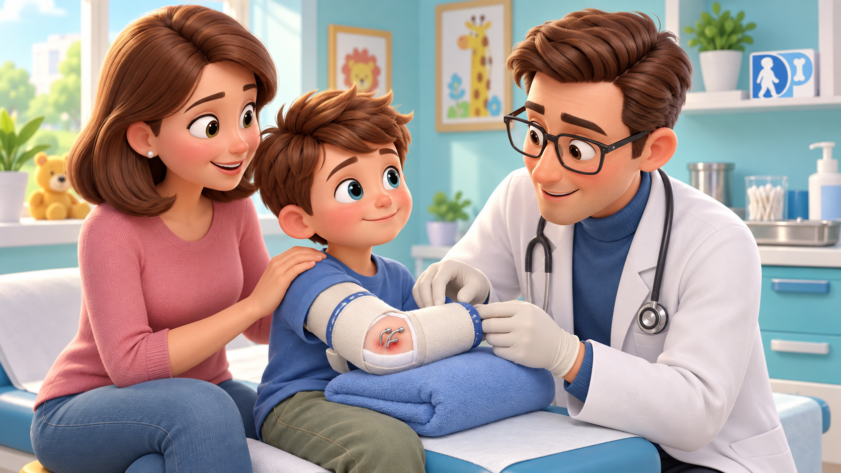
Kirschner wires are used very frequently in paediatric trauma. In the vast majority of children, the course is uneventful: the fracture consolidates, the wires are removed, and that is the end of the matter.
However, an infection can appear at the point where the wire breaches the skin. Most of these infections remain superficial. The real challenge is therefore not to treat every area of erythema as osteomyelitis, but rather to identify the child in whom the infection is no longer following the course of a simple superficial infection.
It is at this transition that one cannot afford to lose time.
An erythematous pin site does not automatically mean the operating theatre
Limited erythema around the wire, mild superficial discharge, and the absence of deep pain or systemic symptoms can be consistent with a superficial pin-site infection.
In such cases, the paediatric literature does not support the need to take the child to the operating theatre automatically. Local wound care and oral antibiotics represent a strategy used in superficial forms, with the antibiotic choice guided by the clinical picture, local epidemiology, and, when indicated and obtainable, microbiology. [2,3]
Nor does the presence of the wire necessarily mean it must be removed immediately. In a paediatric series dedicated to K-wire infections, the authors highlighted that a wire that is still necessary to maintain an ununited fracture can be temporarily retained if the infection is genuinely superficial. [2]
Conversely, if the fracture is already sufficiently stable, wire removal can more readily form part of infection management. However, there is no single rule that applies to every fracture: removing hardware that is still required for stability too early can create a secondary problem.
The crucial aspect is close follow-up. Oral antibiotic therapy should not become a way of postponing the re-evaluation of a condition that continues to deteriorate.
If it does not improve, the clinical reasoning must change
A 2015 paediatric study described 12 children admitted for Kirschner wire-related infection. Clinical presentations ranged from cellulitis to abscess, through to osteomyelitis and one case of toxic shock syndrome. Five children with an abscess, septic arthritis, or osteomyelitis required repeat surgery. A particularly interesting finding was that osteomyelitis was frequently preceded by discharge from the pin site and failure of oral antibiotic therapy. [2]
This does not mean that oral antibiotics are an incorrect treatment. It means that a lack of response to antibiotics is clinical information in itself.
A more recent paper on infectious complications of K-wires suggests, in apparently superficial infections, re-evaluation as early as approximately 48 hours after initiating therapy. If no evident improvement occurs within 48–72 hours, or if the clinical picture worsens, the suspicion of a deeper infection rises and it becomes reasonable to re-evaluate the need for imaging, cultures, drainage, wire removal, or surgical management. [3]
I would not turn these 48 hours into a rigid threshold. A child with fever, significant pain, progressive swelling, marked purulent discharge, or restricted joint range of motion should not have to wait for a timer to run out. Conversely, minor erythema that is rapidly resolving is a completely different issue.
The useful principle is to observe the trajectory of the clinical picture.
When the infection reaches the bone
The new study by Lao and colleagues, published in 2026 in the Journal of Pediatric Orthopaedics, begins precisely from cases in which this transition had already occurred. The authors gathered 16 children with K-wire-related osteomyelitis following the treatment of a closed fracture. Fifteen were managed surgically and one without surgery. All achieved resolution of the infection without recurrence at final follow-up. [1]
The most interesting finding, however, is not the cure rate. It is how the infection behaved.
Osteomyelitis tended to develop along the tract of one of the wires. When that tract crossed important structures, the infection could involve them: in the series, septic arthritis was present in 11 of the 15 evaluable patients, necrosis of a secondary ossification centre in 6 of 10, and partial or complete premature physeal closure in 9 of 15. [1]
These figures must be read with great care. They do not represent the risk of these complications in any child treated with Kirschner wires. The study selected exclusively children who had already developed osteomyelitis. From this cohort, we do not know the denominator of all fractures treated with K-wires, and we therefore cannot deduce the incidence of osteomyelitis, septic arthritis, or physeal damage following standard percutaneous fixation.
They do serve, however, to explain why, when a concrete suspicion of deep infection exists, simply waiting and repeating courses of antibiotics can be the wrong choice.
Superficial and deep are two distinct problems
A superficial pin-site infection and osteomyelitis along the wire tract are not two interchangeable degrees of the same condition.
In the former, local management associated with oral antibiotic therapy—when indicated—and close monitoring may suffice. In the latter, the extent of the infection, fracture stability, potential joint or physeal involvement, and the need for surgical debridement combined with targeted antibiotic therapy all come into play. [1–3]
The series by Lao does not prove that the technique used by the authors—extensive debridement along the wire tract combined with an antibiotic-impregnated bone substitute—is superior to other strategies. It is a retrospective study of only 16 patients, collected at a single centre over a twelve-year period, with no control group. Nevertheless, it is difficult to overlook the anatomical message: once infection has entered the bone, the wire tract can become the route through which it reaches deeper structures. [1]
For this reason, after fixation with Kirschner wires, one should not be alarmed by every minor area of erythema. Nor, however, should one normalise persistent discharge, increasing pain, or a child who is deteriorating rather than improving.
The crucial moment is not when every pin-site infection must be operated on. It is when one must recognise that it is no longer merely a pin-site infection.
References
[1] Lao J, Bai H, Lu S, Li G, Ali S, Zhu T, Liu T, Wang E. Management and Outcome of Kirschner Wire-related Osteomyelitis in Pediatric Patients After Treatment of Closed Fracture. J Pediatr Orthop. Published online September 10, 2026. doi:10.1097/BPO.0000000000003463. PMID: 42720603.
[2] Tosti R, Foroohar A, Pizzutillo PD, Herman MJ. Kirschner wire infections in pediatric orthopaedic surgery. J Pediatr Orthop. 2015;35(1):69-73. doi:10.1097/BPO.0000000000000208. PMID: 24787310.
[3] Infectious complications of K-wire fixation in pediatric fractures: Risk factors and management at a tertiary care center. Injury. 2025. doi:10.1016/j.injury.2025.112944.
Disclaimer: this content is for general informational purposes only. It does not replace an individual medical assessment.
