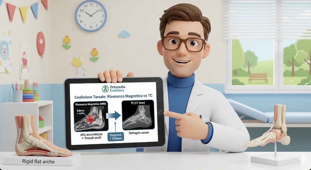
In the clinic, a similar scenario often unfolds: "He's always had somewhat flat feet, but now he's complaining. And he frequently sprains his ankle." Or: "He can no longer play sports like before; he says his ankle is stiff." When hindfoot pain is associated with reduced subtalar joint mobility, tarsal coalition quickly becomes one of the primary hypotheses.
For years, in daily practice, CT scans were considered the "safest" examination to confirm the diagnosis: they clearly define bony bridges and clarify the anatomy. The problem is twofold. First: not all coalitions are fully ossified, especially in the early stages. Second: in children and adolescents, the dominant symptom is often pain, and pain does not only depend on the "presence" of the coalition but also on associated phenomena (bone edema, overload reactions, impingement, soft tissue suffering). Hence the concrete question: can magnetic resonance imaging be as reliable as CT?
A recent study published in European Radiology addressed precisely this point, measuring the diagnostic accuracy of magnetic resonance imaging in recognizing tarsal coalitions. The authors report a cumulative sensitivity of 95.8% and a specificity of 94.3%, with almost perfect agreement among different operators. [1] These numbers, despite the inevitable limitations of a retrospective study, raise the bar: MRI is not just a "useful if you want to see something else" examination, but a test with high performance even for the diagnosis itself.
The real practical advantage, however, is not just the confirmation of the coalition. It's the ability to obtain, with a single examination, two pieces of information that are often both needed: the presence (or absence) of the coalition and the associated findings that help explain the symptoms. [1] In real life, this can make a difference, especially in cases where the clinical picture is "pain-driven" rather than "deformity-driven": children with repeated sprains, pain in the sinus tarsi or hindfoot, functional limitation during sports, and inconclusive X-rays.
This does not mean that CT scans will disappear. There are still situations where millimeter-level bone definition is useful, for example, in preoperative planning or when MRI is not accessible within a reasonable timeframe. The message, however, is clear: in cases of suspected tarsal coalition, magnetic resonance imaging can be considered a first-choice examination because it is accurate and, at the same time, "broadens" the clinical assessment without radiation exposure. [1]
Disclaimer
This content is for informational purposes only and does not replace a medical evaluation. In the presence of persistent pain, limping, foot stiffness, or repeated sprains, a specialist visit is advisable.
References
[1] Marth AA, et al. How accurate is MRI for diagnosing tarsal coalitions? A retrospective diagnostic accuracy study. European Radiology. 2024. PMID: 37855854.
PubMed: https://pubmed.ncbi.nlm.nih.gov/37855854/
Full text (PMC): https://pmc.ncbi.nlm.nih.gov/articles/PMC11126476/
