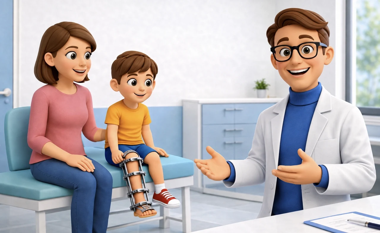
Congenital pseudarthrosis of the tibia (CPT) remains one of the most complex conditions in pediatric orthopedics. Treatment must achieve consolidation, but also reduce the risk of refracture and manage long-term deformity, ankle instability, and limb length discrepancy.
A 2025 review in JBJS Reviews summarizes the current approach, where the biology of the pseudarthrosis site and mechanical stability are addressed together. [1]
Biology and Mechanics
CPT is strongly associated with neurofibromatosis type 1 and an unfavorable biological environment, with hamartomatous tissue and cellular alterations that hinder healing. [1]
Resection of pathological tissue, bone grafting, and stable mechanical support are therefore combined in the main reconstructive protocols. Classifications such as Andersen, Boyd, Crawford, and Paley are useful for describing the condition, but do not solely determine the strategy.
Refracture After Union
Refracture is one of the central problems in follow-up. A bone that consolidates with a reduced load-bearing cross-section can remain vulnerable even after radiographic union.
Paley emphasized the role of biomechanical reconstruction and increasing the load-bearing cross-section of the limb as part of refracture prevention. [2]
Tibia-Fibula Cross-Union
Cross-union deliberately creates a bone bridge between the tibia and fibula, increasing the overall cross-section of the reconstructed segment.
A retrospective series of 39 pseudarthroses in 36 patients described a protocol involving hamartoma resection, grafting, intramedullary support, and cross-union. In the reported cohort, union was achieved in all cases, and no refractures were observed during the available follow-up, up to approximately seven years. [3]
These are significant results, but they come from highly experienced centers and non-randomized studies. The technique requires complex planning and cannot be automatically considered reproducible in every context. [1][3]
Intramedullary Support and External Fixation
Intramedullary nails, including telescopic systems like the Fassier-Duval, can maintain mechanical protection during growth. In more complex protocols, they are combined with grafting and, in some cases, external fixation.
The circular fixator maintains a role when it is necessary to correct deformities, manage shortening, or control the axis. Vascularized fibular graft represents another option historically used in selected cases. [1]
Growth often necessitates revisions and subsequent procedures. For example, problems with migration or embedment of telescopic nails have been described, which can complicate planned exchanges. [3][4]
Biological Adjuvants
BMP and bisphosphonates have been used as adjuvants in some protocols. The 2025 review still considers the evidence for their independent contribution to be inconsistent. [1]
They do not replace adequate resection, grafting, and stability. Their potential role should be interpreted within complex reconstructive strategies, especially in patients with NF1.
Sequelae During Growth
Even after stable union, limb length discrepancy, axial deformity, ankle alterations, and the need for further interventions may remain. For this reason, the outcome of CPT is not measured solely at the time of initial consolidation.
Follow-up must accompany growth and consider the axis, length, stability, and function of the entire limb.
Limitations of Evidence
CPT is rare, and much of the literature comes from retrospective series from specialized centers. Directly comparing different techniques is difficult because severity, age, biological protocols, and fixation methods vary. [1][3]
The series on cross-union are promising, especially for refracture control, but independent confirmations and very long follow-ups are needed.
Disclaimer
This content is for informational purposes only and does not replace an individual clinical evaluation.
References
[1] Al Ramlawi A, Chenard SW, Sidani M, Herzenberg JE, Schoenecker JG, McClure PK. Congenital Pseudarthrosis of the Tibia: A Comprehensive Literature Review. JBJS Rev. 2025;13(6). doi:10.2106/JBJS.RVW.25.00035. PMID: 40472164.
PubMed: https://pubmed.ncbi.nlm.nih.gov/40472164/
[2] Paley D. Congenital pseudarthrosis of the tibia: biological and biomechanical considerations to achieve union and prevent refracture. J Child Orthop. 2019;13(2):120-133. doi:10.1302/1863-2548.13.180147. PMID: 30996736. PMCID: PMC6442511.
PubMed: https://pubmed.ncbi.nlm.nih.gov/30996736/
PMC full text: https://pmc.ncbi.nlm.nih.gov/articles/PMC6442511/
[3] Paley D, et al. Cross-Union Surgery for Congenital Pseudarthrosis of the Tibia. PMID: 34202921. PMCID: PMC8303361.
PubMed: https://pubmed.ncbi.nlm.nih.gov/34202921/
PMC full text: https://pmc.ncbi.nlm.nih.gov/articles/PMC8303361/
[4] McClure PK, Franzone JM, Herzenberg JE. Challenges with Fassier-Duval rod exchanges in congenital pseudarthrosis of the tibia: explant roadblock and solution. J Pediatr Orthop B. 2022;31(1):e95-e100. doi:10.1097/BPB.0000000000000907. PMID: 34380988.
