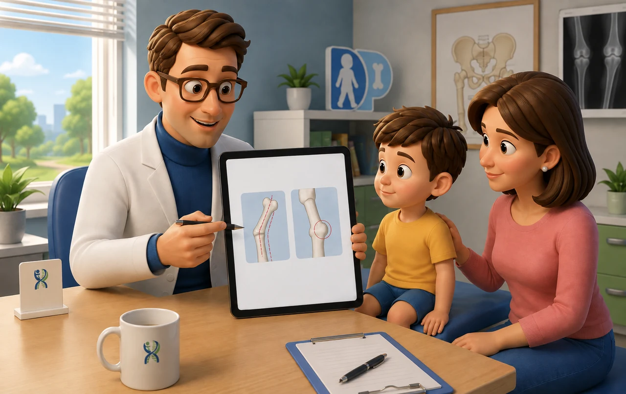
After cast removal, a child’s arm or leg may not look the way the parent expected. A hard prominence can be felt at the fracture site, sometimes quite clearly; alternatively, the X-ray may still show an angulated bone that does not appear completely straight.
Both findings can be compatible with normal healing. The prominence is often fracture callus, the new bone produced during healing. Residual angulation, on the other hand, relates to remodeling, the process through which a growing bone can gradually change its shape even after the fracture has healed. These are two different phenomena, and they occur over different timescales. [1,2]
The bump after a fracture
During healing, the body produces new bone tissue around the fracture. The callus may be abundant, and in more superficial bones it can be easy to see or feel. This often happens, for example, in the forearm or clavicle.
During the first few weeks, the prominence may even seem larger than expected. The bone is then gradually reorganized: part of the callus is resorbed, and its structure changes over time. A hard, painless prominence located exactly where the fracture occurred does not, by itself, mean that the bone has healed incorrectly. [1]
The X-ray raises a somewhat different issue. In a child, a fracture may be clinically healed even if the image does not yet show a perfectly aligned bone. Some residual angulation may correct with growth, but how much correction can occur depends greatly on the individual fracture. [1,2]
Age is important: all other factors being equal, a younger child has greater remodeling potential than an adolescent approaching skeletal maturity. The location of the fracture also matters. Deformities close to a highly active growth plate tend to remodel better than those in the middle portion of a bone. The plane of the angulation in relation to the movement of the nearby joint also affects the potential for correction. [1,2]
For this reason, there is no single number of degrees that can be considered acceptable for every pediatric fracture. An X-ray that looks markedly angulated in a young child may still have a favorable outcome; a less obvious deformity in another location or at another age may be less acceptable.
Not everything straightens with growth
The remodeling capacity of a child’s bone is remarkable, but it has clear limits. Rotational deformities are an important example: if a bone heals while rotated around its own axis, the ability to correct that rotation spontaneously is much more limited than the ability to correct many angular deformities. [2]
This is also why some fractures can be allowed to heal with a certain degree of residual angulation, while others require another reduction or surgical stabilization. The aim is not to pursue an aesthetically perfect X-ray in every child, but to predict what that specific deformity is likely to do during growth.
The timing of fracture healing and the timing of remodeling are also different. When the cast is removed, the fracture has reached sufficient stability, but the bone can continue to change for months and, in children with substantial growth remaining, sometimes for even longer. [1]
When another check is advisable
A hard bump that is not painful and corresponds exactly to the fracture site may therefore be normal. The situation is different if pain is increasing, new swelling or redness develops, the child loses a function that had already returned, or the deformity appears to be getting worse.
Persistent limitation of joint motion and an obvious rotational abnormality also deserve reassessment. In these situations, it is not advisable simply to wait for growth to correct everything, because different deformities do not have the same remodeling potential.
After a fracture, therefore, seeing a residual bump or some angulation does not automatically mean that treatment has failed. To understand whether that appearance is likely to improve, it is necessary to consider the child’s age, the fracture location, the type of deformity, and the amount of growth remaining.
References
[1] Gamble JG. Pediatric Fracture Remodeling: From Wolff to Wnt. Cureus. 2025;17(1):e78266. PMID: 39897217. PMCID: PMC11782688. doi:10.7759/cureus.78266.
[2] Naik P. Remodelling in Children’s Fractures and Limits of Acceptability. Indian J Orthop. 2021;55(3):549–559. PMID: 33995859. PMCID: PMC8081818. doi:10.1007/s43465-020-00320-2.
Disclaimer: This content is intended for general informational purposes and does not replace an individual medical assessment.
