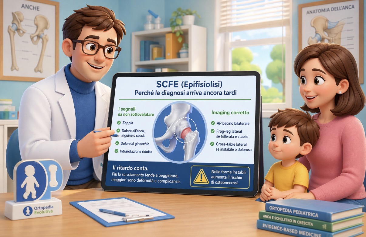
SCFE (Slipped Capital Femoral Epiphysis) is a diagnosis that carries significant weight: because it is common in practice, because it can present deceptively, and because diagnostic delay has concrete consequences. It's not just "lost time"; it's time during which the slip can worsen, residual deformity increases, and in unstable forms, the risk of the most feared complications grows [1–6].
The first deception: the knee
One of the most classic mechanisms of delayed diagnosis is presentation with knee pain. In these cases, the diagnostic chain tends to remain "stuck" on the wrong area: the knee is examined, knee imaging is requested, rest is advised... and the hip remains outside the reasoning.
The most convincing epidemiological data comes from a national cohort using primary care databases: when the onset is in the knee, the diagnostic delay is significantly greater compared to cases with hip/groin pain or limping as the main symptom [5].
In practice, the simplest (and most effective) rule is this: in an adolescent with limping and knee pain, one must always "go up" to the hip, evaluating rotation and the presence of obligatory external rotation in flexion (Drehmann's sign) [1–3].
The second deception: incomplete or "unfocused" imaging
The problem is not "the frog leg view is missing"; the problem is that often the right projection is missing.
In suspected cases, imaging must include projections that make the slip visible on the sagittal plane. In stable forms, the combination of AP pelvis + frog-leg lateral projection is generally the most useful; in unstable forms (or when the patient cannot tolerate the position), a cross-table lateral is preferable to avoid painful and potentially risky maneuvers [1–3].
An important aspect is that "classic" signs (such as Klein's/Trethowan's line) may be insensitive in the early stages. For this reason, many summaries suggest also reasoning with more robust measurements (e.g., Southwick angle) and with systematic comparison to the contralateral side, especially when the clinical picture is suggestive but the X-ray is not striking [1–3].
Why delay matters: deformity and complications
Saying that "SCFE worsens over time" is not a generic phrase; it is a clinical concept. When diagnosis is delayed, the slip tends to progress, residual deformity increases, and the risk profile changes in the medium to long term (femoroacetabular impingement, pain, functional limitation, early osteoarthritis) [1–3].
In unstable forms, the main problem is osteonecrosis (AVN). A 2025 meta-analysis of 688 hips estimates an AVN incidence of around 23% in unstable cases and identifies male gender, moderate-to-severe slip, acute presentation, and—a delicate issue—reduction attempts, particularly closed reduction, as factors associated with increased risk [6]. This should be read with caution (retrospective data, confounding by severity), but it is useful to avoid "simplistic" interpretations such as "always reduce, no matter what."
Why diagnosis is still missed (and it's not one person's fault)
A 2023 review dedicated to diagnostic errors in SCFE brings together very concrete causes: underestimated initial symptoms, first contact with non-orthopedic professionals, imaging requested for the wrong area, suboptimal radiographic interpretation in early forms, and variable clinical presentations [4].
The message that emerges, in my opinion, is the right one: the error is often in the process, not in individual competence. And processes can be improved.
A practical approach that reduces delays
Without wanting to list commands, there are three points that, if applied consistently, truly change the frequency of delayed diagnoses:
- "High" clinical suspicion in the right patient: adolescent with limping, hip/groin/thigh pain, or knee pain, especially with reduced internal rotation [1–3][5].
- Targeted imaging: bilateral pelvis/hips with adequate AP and lateral views (frog-leg if tolerated and stable; cross-table if unstable or not tolerated) [1–3].
- Do not dismiss convincing clinical findings too quickly: if the history and physical examination "speak," a single non-definitive X-ray should not close the case; close re-evaluation is needed, and in selected cases, advanced imaging [3][4]. (This is not a single recipe; it is a principle of diagnostic safety.)
In summary
SCFE continues to be diagnosed late, especially when it presents "sideways": knee pain, vague symptoms, incomplete imaging. Recent literature reinforces a simple point: the best way to reduce complications is to reduce delays, and delays are reduced with clinical suspicion, adequate projections, and shared pathways [1–6].
Disclaimer
This content is for informational purposes only and does not replace an individual clinical evaluation.
References
[1] Webb CW, Liu R, Bouchereau-Lal N. Slipped Capital Femoral Epiphysis: Rapid Evidence Review. American Family Physician. 2025;112(4):414-423.
https://www.aafp.org/pubs/afp/issues/2025/1000/slipped-capital-femoral-epiphysis.html
[2] POSNA Study Guide – SCFE.
https://posna.org/physician-education/study-guide/scfe-%28slipped-capital-femoral-epiphysis%29
[3] Johns K, Mabrouk A, Tavarez MM. Slipped Capital Femoral Epiphysis. StatPearls. Updated 2023.
https://www.ncbi.nlm.nih.gov/books/NBK538302/
[4] Diagnosis of Slipped Capital Femoral Epiphysis: How to Stay out of Trouble? Children (Basel). 2023;10(5):778.
https://www.mdpi.com/2227-9067/10/5/778
[5] Perry DC, Metcalfe D, Costa ML, Van Staa T. Presentation and delay in diagnosis of slipped capital femoral epiphysis: a nationwide cohort study. Arch Dis Child. 2017. (full text)
https://pmc.ncbi.nlm.nih.gov/articles/PMC5754864/
[6] Xu Z, Zhu L, Kong L, et al. Risk factors associated with avascular necrosis following unstable slipped capital femoral epiphysis: a systematic review and meta-analysis. PLOS ONE. 2025;20(7):e0329275.
