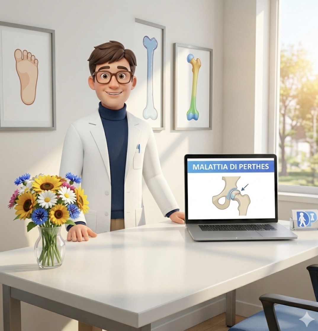
Legg-Calvé-Perthes disease is one of those conditions that seems to have been known forever, yet continues to defy simplification. We often describe it as idiopathic osteonecrosis of the femoral head in children, with a fairly recognizable sequence: necrosis, revascularization, resorption, reossification, and healing. So far, so clear. The problem is that between the beginning and the healing, the most difficult part plays out: understanding which femoral head will heal spherical and congruent, and which will leave a deformity capable of affecting the hip for life [1].
The review by Gilbert, Laine, Martin, Sankar, and Kim, published in the Journal of the American Academy of Orthopaedic Surgeons, has the merit of not offering an easy answer. It reminds us that the natural history is often favorable in younger children, especially under 6 years old, while it becomes more concerning in older children, particularly over 8–10 years old. But it doesn't turn these thresholds into a rigid algorithm. In Perthes, age matters a lot, but it's not the only variable: the stage of the disease, hip mobility, the extent of epiphyseal involvement, congruence with the acetabulum, and the ability to limit mechanical damage while the head is biologically fragile all matter [1].
The historical concept of containment remains central. The goal is to keep the femoral head as well-centered and congruent as possible within the acetabulum while it goes through the revascularization and remodeling phase. Many strategies stem from this: maintaining range of motion, reducing load during symptomatic periods, using exercises, braces, or casts in selected cases, and in higher-risk patients, considering femoral osteotomies, pelvic osteotomies, or combined procedures [1].
The point, however, is that saying "containment" doesn't mean the decision has been made. A recent surgical review of options in Perthes highlights just how difficult it is to demonstrate the clear superiority of a single surgical strategy, because the disease is relatively rare, has a variable natural history, and requires many years to understand the final outcome [3]. In other words: it's not enough to choose a "correct" technique; you have to choose the right patient at the right time.
Here, the analysis published in 2026 in the Bone & Joint Journal, based on data from a large prospective multicenter study and a modern measure of sphericity, the sphericity deviation score, also becomes interesting. The authors report that, in patients with a bone age over 6 years, proximal femoral osteotomy or Salter osteotomy were associated with better sphericity scores compared to no treatment or mobility exercises alone. Furthermore, in the regression tree analysis, patients with a chronological age over 7.6 years treated with femoral osteotomy had better outcomes than those not treated surgically [2]. This is not a license to operate on everyone, but it reinforces the idea that in older children and higher-risk cases, surgical containment still has a strong rationale.
In the clinic, the most challenging case is almost never the very young child with mild symptoms and good mobility. It's the 7–8-year-old child, perhaps arriving after weeks of intermittent limping, with reduced abduction and internal rotation, an already quite eloquent X-ray, but a family convinced that "it will pass like synovitis." At that moment, the temptation can be twofold: to reassure too much, because many Perthes cases improve, or to be too aggressive too soon, because the X-ray is frightening. Recent literature encourages staying in the middle: reading the risk, not just the image.
The most modern part of the JAAOS review, in my opinion, is precisely this: Perthes is no longer described solely as a mechanical issue. Containment remains fundamental, but recent research is increasingly focusing on imaging predictors and the possibility of understanding, and perhaps modifying, the biological processes that follow the initial vascular insult [1]. A 2025 review in Frontiers in Physiology also moves in this direction, discussing the disease from a molecular and cellular biology perspective: not yet something that will change tomorrow's visit, but a clear signal that the future of Perthes will not be made only of osteotomies and braces [4].
This is also important for communicating with non-pediatric colleagues. Perthes is not a "long synovitis" nor is it a diagnosis to be filed away with a single initial X-ray if the clinical picture doesn't fit. A child with persistent limping, hip or knee pain, limited abduction or internal rotation deserves a careful hip evaluation. The sooner the problem is identified, the more realistic it is to preserve mobility, containment, and the possibility of remodeling.
The practical message I take home is not that we have a new definitive answer today. It's almost the opposite. After more than a hundred years, Perthes remains a disease where clinical experience matters a lot, but it must be nourished by better data: more predictive imaging, more refined measures of the final femoral head shape, long follow-ups, and multicenter studies. The choice between observation, physiotherapy, load limitation, bracing, osteotomy, or joint distraction cannot be reduced to a single table [1–3].
If I had to summarize the meaning of the update in one sentence, I would say this: in Perthes disease, containment remains a key word, but today the real goal is to better understand who needs containment, when, and at what biological and functional cost.
Disclaimer
This content is for informational purposes only and does not replace individual clinical evaluation.
References
[1] Gilbert SR, Laine JC, Martin BD, Sankar WN, Kim HKW. Legg-Calvé-Perthes Disease. J Am Acad Orthop Surg. 2026;34(4):e488-e497. Epub 2025 Sep 24. doi:10.5435/JAAOS-D-24-01469. PMID: 40991851.
PubMed: https://pubmed.ncbi.nlm.nih.gov/40991851/
[2] Herring JA, Kim HKW, et al. The outcomes of treatment and prognostic determinants of patients with Perthes’ disease: reanalyses of large prospective multicentre study data using the sphericity deviation score. Bone Joint J. 2026. PMID: 41619794.
PubMed: https://pubmed.ncbi.nlm.nih.gov/41619794/
[3] Braun S, Adolf S, Brenneis M, Boettner F, Meurer A, et al. Legg-Calvé-Perthes disease – surgical treatment options. Arch Orthop Trauma Surg. 2025;145:186. doi:10.1007/s00402-025-05801-3.
Journal: https://link.springer.com/article/10.1007/s00402-025-05801-3
[4] Zheng X, Dong Z, Ding X, Huang Q, Tang S, Zhang Y, Li B, Liao S. Progress in understanding Legg–Calvé–Perthes disease etiology from a molecular and cellular biology perspective. Front Physiol. 2025;16:1514302. doi:10.3389/fphys.2025.1514302.
Journal: https://www.frontiersin.org/journals/physiology/articles/10.3389/fphys.2025.1514302/full
