Paediatric Orthopaedic Specialist
Köhler disease (active management)
Home Exercise Program
Not every exercise listed here is suitable for every child: the selection is individual and is defined during the consultation.
For whom: After diagnosis of Köhler disease. Respect offloading or immobilisation; introduce standing exercises only with authorised weight bearing and no midfoot pain.
Dose and frequency: Perform only the exercises selected during the visit. The listed dosages are examples to adapt: follow the dose, frequency, rest and progression prescribed for age, symptoms and phase. Do not automatically perform the whole list.
Equipment and supervision: A stable, clear surface, comfortable clothing and a secure support. Check resistance-band anchors. Adult supervision; avoid marbles and small objects if the child may put them in their mouth, using a towel instead.
Avoid: Forcing a painful or unauthorised movement; Increasing load, resistance or frequency without the agreed rest periods; Doing balance exercises without stable support or supervision.
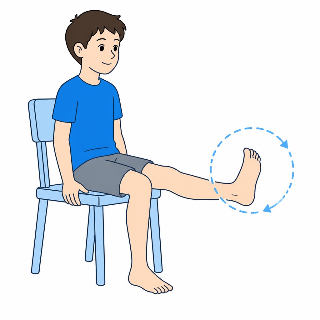
1.Non-weight bearing foot mobility (alphabet)
Maintains mobility without loading the painful navicular
Seated with foot raised. "Write" alphabet letters with toe tip. This gently moves all foot joints without loading the navicular. Add ankle circles in both directions. All movements should be pain-free.
Full alphabet + 10 circles each direction, 2-3 times daily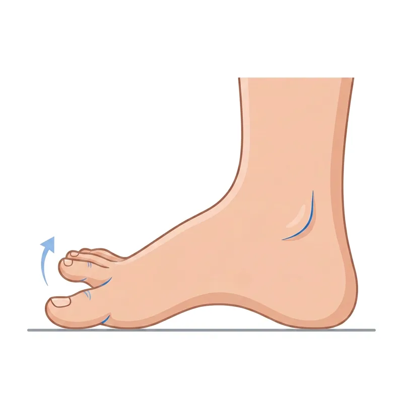
2.Toe yoga and toe control (seated)
Strengthens intrinsic foot muscles without weight on navicular
SEATED (to avoid foot loading). Alternate: lift big toe keeping others down, then reverse. Curl toes as if grasping, then fully extend. Pick up small objects with toes. These exercises keep foot muscles active during the relative rest period.
10-15 reps per movement, 1-2 times daily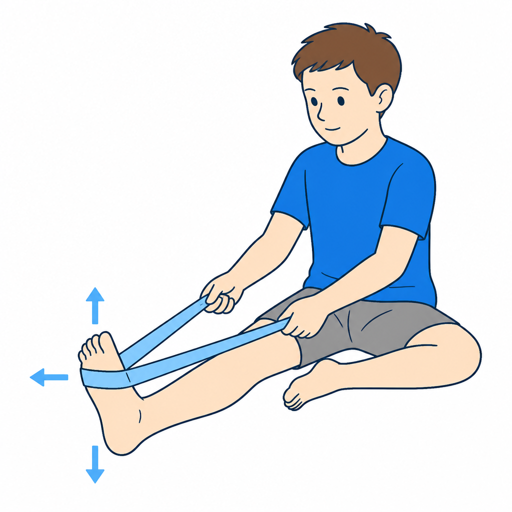
3.Ankle strengthening with band (seated)
Maintains ankle strength during rest phase
Seated with band around foot. Perform 4 movements against resistance: dorsiflexion (up), plantarflexion (down), inversion (in), eversion (out). Use light resistance. This maintains muscle strength without foot loading.
3 × 12-15 each direction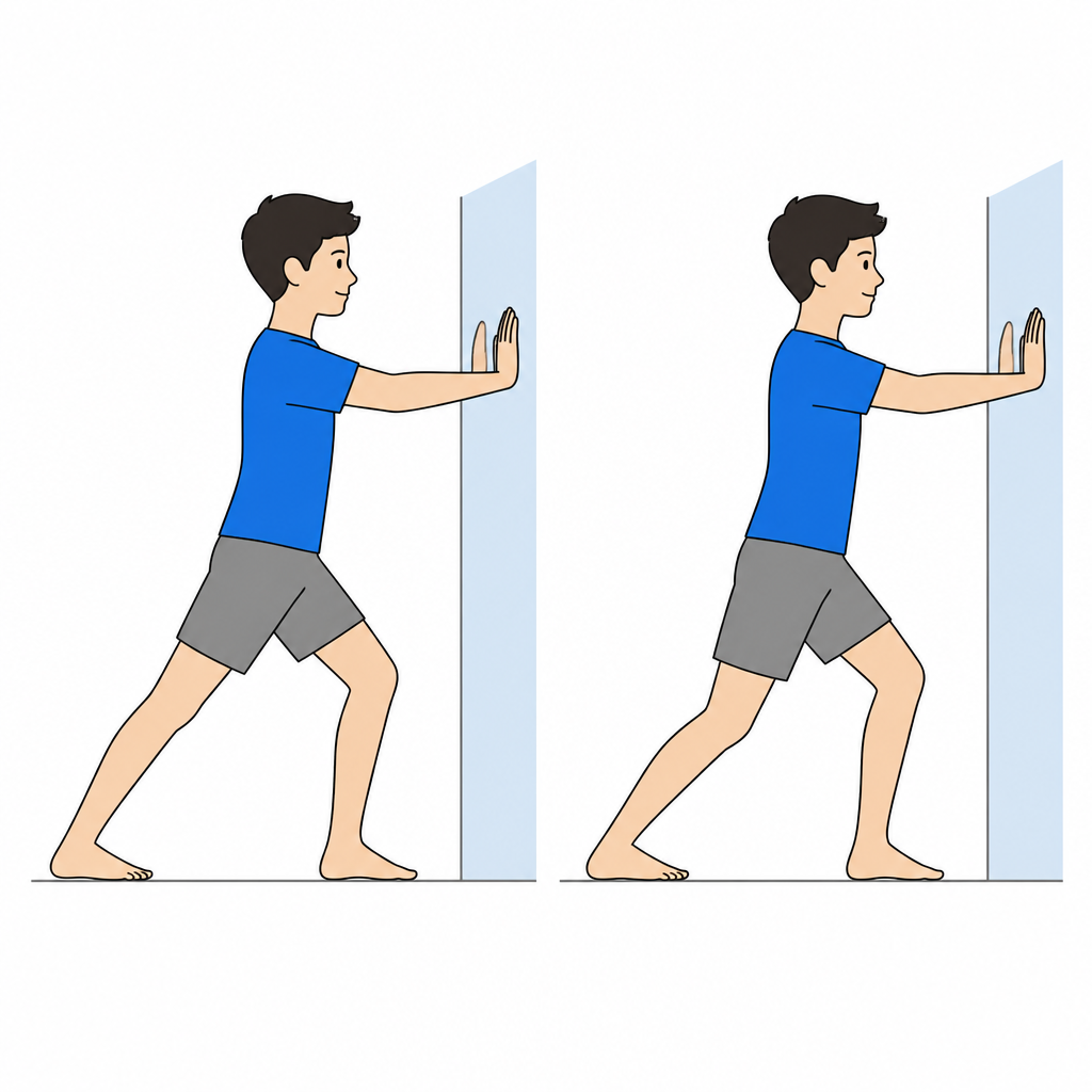
4.Calf stretch (gentle)
Reduces stiffness that increases midfoot loading
Facing a wall, leg behind with the heel down, knee straight and then bent. Push the hips forward until a gentle lengthening is felt. In Köhler disease the goal is to preserve ankle mobility during the reduced-loading period: the stretch must always be pain-free.
3 × 20-30 seconds per position, 1-2 times daily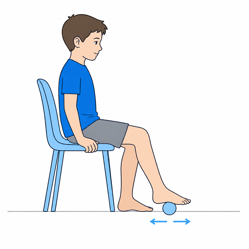
5.Plantar massage with a ball
Relaxes plantar muscles while avoiding the navicular area
Seated, roll a soft ball under the sole focusing on the heel and forefoot while AVOIDING direct pressure on the inner midfoot (where the navicular lies). Light pressure, never painful. Useful before mobility exercises.
2-3 minutes, once daily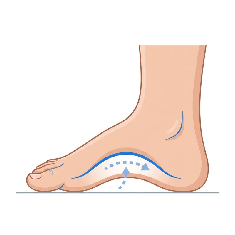
6.Progressive short-foot
Reactivates the intrinsic muscles supporting the arch, first unloaded then loaded
PHASE 1 (seated, no weight): place the foot on the floor and "shorten" it by lifting the arch without curling the toes. Hold 5 seconds. PHASE 2 (when pain-free, standing on both legs): same movement with reduced load. Progress only if no pain or limping appears the next day.
10 reps × 5 seconds, twice daily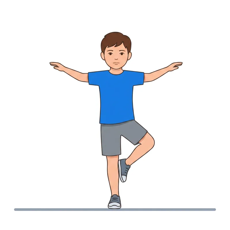
7.Balance and proprioception (advanced phase)
Restores foot control before full return to activity
After clearance for weight bearing, start on firm ground with eyes open, next to a secure support and with adult supervision. Place the other foot down before losing balance. Progress to an unstable surface or eyes-closed work only under direct professional supervision; do not add these challenges independently. Stop for pain or instability.
3 × 20-30 seconds per leg, once daily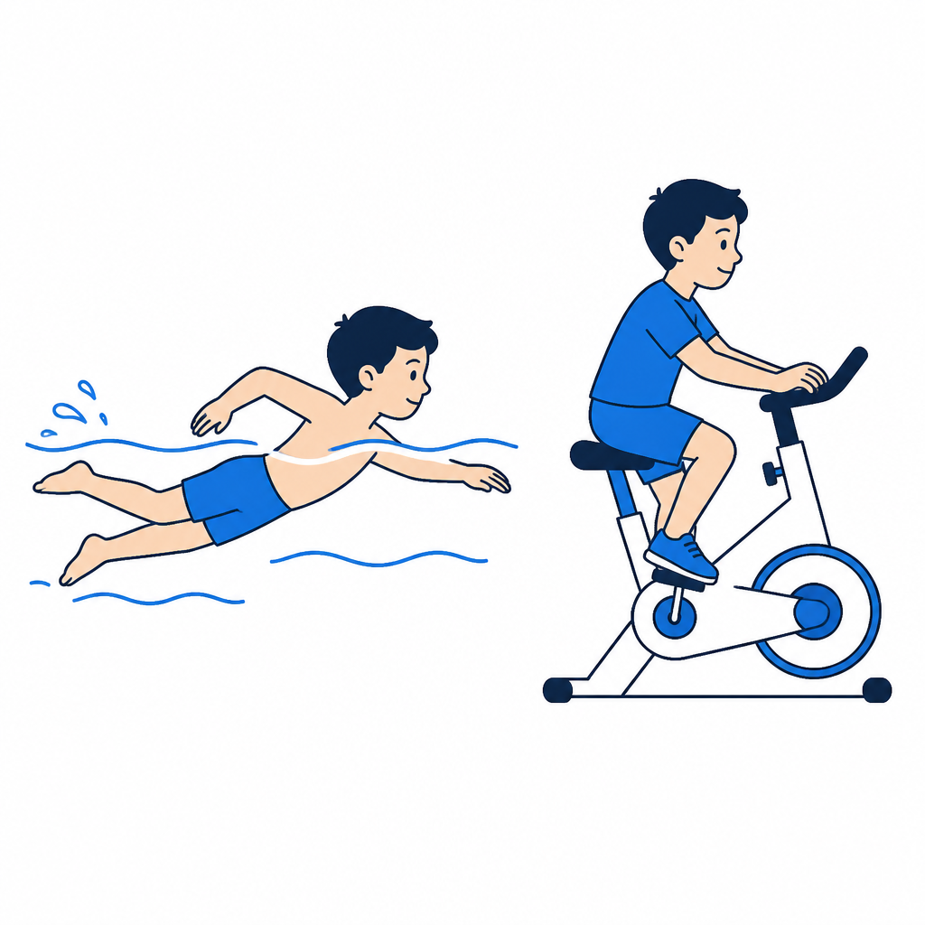
8.Low-impact activities
Maintains fitness without loading the navicular during the painful phase
During the symptomatic phase, keep exercising with swimming and cycling (high saddle, midfoot pedalling, no standing on the pedals). These train heart and muscles without foot impact. Avoid running, jumping and team sports until the specialist advises otherwise.
20-30 minutes, 2-3 times weekly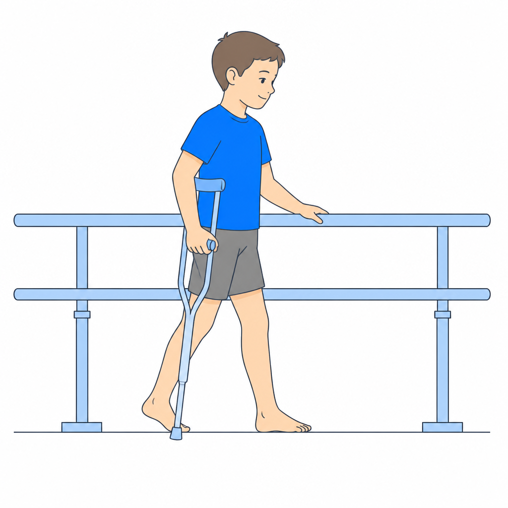
9.Gradual return to weight bearing
Protected progression toward activity resumption
When pain significantly reduces: 1) Walking with cushioning shoes and supportive insole. 2) Walking on flat surfaces, short distances. 3) Gradual distance increase. 4) Light activities (swimming, cycling). 5) Light running only when completely pain-free. Return to sport follows specialist guidance.
Individualized progression, guidance during visit
RED FLAGS - When to stop and contact the doctor immediately
Stop the exercises immediately and contact the specialist (or go to the emergency department if severe) if any of the following occur:
- Acute or sudden pain during exercise
- Significant swelling after exercises
- Pain that progressively worsens over days
- Fever or general malaise
- Difficulty walking or using the limb after exercises
- Any neurological symptoms (tingling, numbness, sudden weakness)
- A red or hot joint, or inability to bear weight: seek prompt assessment; use emergency care if severe.
In case of doubt, always prefer a check-up over continuing the exercises.
Dott. Daniele Priano
Paediatric Orthopaedics - ASST Gaetano Pini-CTO
Content version: 9 September 2026
ortopediaevolutiva.com • Printed: 13/09/2026
 Web version
 Google
 MioDottore
Prepare the visit handout
Use the condition and phase indicated by your clinician. You can leave out individual exercises; instructions, illustrations and safety advice remain together.
Exclude exercises if needed
9 of 9 exercise cards included
The full web page remains available. Your selection is not saved or shared.
Before You Start
Who is this guide for?
After diagnosis of Köhler disease. Respect offloading or immobilisation; introduce standing exercises only with authorised weight bearing and no midfoot pain.
What you need
A stable, clear surface, comfortable clothing and a secure support. Check resistance-band anchors. Adult supervision; avoid marbles and small objects if the child may put them in their mouth, using a towel instead.
Suggested frequency
Perform only the exercises selected during the visit. The listed dosages are examples to adapt: follow the dose, frequency, rest and progression prescribed for age, symptoms and phase. Do not automatically perform the whole list.
Common mistakes to avoid
- •Forcing a painful or unauthorised movement
- •Increasing load, resistance or frequency without the agreed rest periods
- •Doing balance exercises without stable support or supervision
⚠️ Red flags — when to stop and contact the doctor immediately
Stop exercises immediately and contact the specialist if any of the following occur:
- Acute or sudden pain during exercise
- Significant swelling after exercises
- Pain that progressively worsens over days
- Fever or general malaise
- Difficulty walking or using the limb after exercises
- Any neurological symptoms (tingling, numbness, sudden weakness)
- A red or hot joint, or inability to bear weight: seek prompt assessment; use emergency care if severe.
Detailed Exercises
- 1
Non-weight bearing foot mobility (alphabet)
Maintains mobility without loading the painful navicular
Seated with foot raised. "Write" alphabet letters with toe tip. This gently moves all foot joints without loading the navicular. Add ankle circles in both directions. All movements should be pain-free.
Full alphabet + 10 circles each direction, 2-3 times daily - 2
Toe yoga and toe control (seated)
Strengthens intrinsic foot muscles without weight on navicular
SEATED (to avoid foot loading). Alternate: lift big toe keeping others down, then reverse. Curl toes as if grasping, then fully extend. Pick up small objects with toes. These exercises keep foot muscles active during the relative rest period.
10-15 reps per movement, 1-2 times daily - 3
Ankle strengthening with band (seated)
Maintains ankle strength during rest phase
Seated with band around foot. Perform 4 movements against resistance: dorsiflexion (up), plantarflexion (down), inversion (in), eversion (out). Use light resistance. This maintains muscle strength without foot loading.
3 × 12-15 each direction - 4
Calf stretch (gentle)
Reduces stiffness that increases midfoot loading
Facing a wall, leg behind with the heel down, knee straight and then bent. Push the hips forward until a gentle lengthening is felt. In Köhler disease the goal is to preserve ankle mobility during the reduced-loading period: the stretch must always be pain-free.
3 × 20-30 seconds per position, 1-2 times daily - 5
Plantar massage with a ball
Relaxes plantar muscles while avoiding the navicular area
Seated, roll a soft ball under the sole focusing on the heel and forefoot while AVOIDING direct pressure on the inner midfoot (where the navicular lies). Light pressure, never painful. Useful before mobility exercises.
2-3 minutes, once daily - 6
Progressive short-foot
Reactivates the intrinsic muscles supporting the arch, first unloaded then loaded
PHASE 1 (seated, no weight): place the foot on the floor and "shorten" it by lifting the arch without curling the toes. Hold 5 seconds. PHASE 2 (when pain-free, standing on both legs): same movement with reduced load. Progress only if no pain or limping appears the next day.
10 reps × 5 seconds, twice daily - 7
Balance and proprioception (advanced phase)
Restores foot control before full return to activity
After clearance for weight bearing, start on firm ground with eyes open, next to a secure support and with adult supervision. Place the other foot down before losing balance. Progress to an unstable surface or eyes-closed work only under direct professional supervision; do not add these challenges independently. Stop for pain or instability.
3 × 20-30 seconds per leg, once daily - 8
Low-impact activities
Maintains fitness without loading the navicular during the painful phase
During the symptomatic phase, keep exercising with swimming and cycling (high saddle, midfoot pedalling, no standing on the pedals). These train heart and muscles without foot impact. Avoid running, jumping and team sports until the specialist advises otherwise.
20-30 minutes, 2-3 times weekly - 9
Gradual return to weight bearing
Protected progression toward activity resumption
When pain significantly reduces: 1) Walking with cushioning shoes and supportive insole. 2) Walking on flat surfaces, short distances. 3) Gradual distance increase. 4) Light activities (swimming, cycling). 5) Light running only when completely pain-free. Return to sport follows specialist guidance.
Individualized progression, guidance during visit
Frequently Asked Questions
Can exercises be done with Köhler disease?
In the acute phase (pain and limping), rest is priority. When pain subsides, non-weight bearing mobility exercises and foot muscle strengthening are useful to maintain function during spontaneous healing. Köhler disease resolves on its own in 1-2 years.