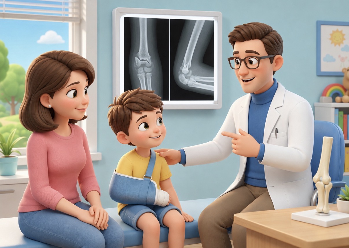
This study addresses a very specific problem: what happens in the days after a child's supracondylar fracture is selected for non-surgical treatment.
The issue is the stability of the reduction. An acceptable post-reduction X-ray does not guarantee that the position will remain unchanged during the first few weeks.
The Clinical Problem
A supracondylar fracture of the humerus is one of the most frequent elbow fractures in children. In some cases, surgical treatment with reduction and Kirschner wires is clearly indicated. In others, especially in less unstable forms, non-surgical treatment with closed reduction and immobilization may be reasonable.
But once the conservative path is chosen, the real question is no longer just: "Is the post-reduction X-ray acceptable?"
The question becomes: Will this reduction remain stable in the coming days?
This is where this study comes in.
The authors retrospectively analyzed 218 children between 3 and 14 years old with Gartland II or selected Gartland III supracondylar humeral fractures, treated non-surgically with closed reduction and custom-molded triplanar immobilization [1].
Type III fractures were not indiscriminately included: they were considered only if, after reduction, they proved stable to fluoroscopic stress testing. During the study period, 42 type III fractures were excluded precisely because they were unstable and converted to surgical treatment [1].
This needs to be stated, but without making it the focus of the commentary. It merely clarifies the scope: we are not talking about all displaced supracondylar fractures, nor an invitation to conservatively treat unstable type III fractures.
When Does Loss of Reduction Occur?
Out of 218 children, 39 experienced radiographic loss of reduction, accounting for 17.9% [1]. By the fourth day, the cumulative incidence of redisplacement was already 6.9%; by the fourteenth day, it reached 15.1%. Approximately 87% of losses of reduction were therefore detected within the first two weeks [1].
The authors describe two critical windows:
- a very early one, around the 3rd-4th day;
- a second one between the 7th and 14th day.
For follow-up, this data is simple to use: earlier check-ups are more likely to detect displacement while it can still be corrected.
Why Exactly the First 14 Days?
The explanation proposed by the authors is plausible.
In the very first days, the inflammatory phase prevails: edema, swelling, tense soft tissues. Stability depends almost entirely on the quality of the reduction and the effectiveness of immobilization.
Between 7 and 14 days, something different happens: the swelling may reduce, the limb may "deflate" inside the splint, but the callus is not yet mature enough to guarantee true biological stability. In that phase, external containment may lose effectiveness before the fracture is stable on its own [1].
This is a sensible biomechanical interpretation, but it should not be sold as absolute certainty. The study is retrospective, and displacements were detected only at scheduled check-ups. Therefore, the peak at days 3-4 and that between days 7 and 14 may partly depend on the X-ray schedule. The authors correctly admit this [1].
I like this detail because it makes the work honest: the message is strong, but it is not forced beyond the data.
Which Fractures Displace More?
The authors found two independent predictive factors for loss of reduction:
- significant initial lateral displacement, measured as LDP >85%;
- severe soft tissue swelling.
In the multivariate model, LDP >85% had a hazard ratio of 3.52, and severe swelling had a hazard ratio of 3.08. Gartland classification, although significant in univariate analysis, lost significance after adjustment [1].
This is a clinically useful point.
Classification is useful, of course. But the child's actual elbow tells more than just the "type II" or "type III" box. What matters is how displaced the fracture was initially, how affected the soft tissues were, how stable it is after reduction, and how reliable the immobilization is.
Classification alone describes only part of the problem. Initial displacement, soft tissues, stability achieved after reduction, and the quality of immobilization also matter.
To Whom Do These Data Apply?
These results do not automatically broaden the indications for conservative treatment and do not concern unstable Gartland III fractures, which were excluded in the study if they failed the fluoroscopic stress test. The useful data concerns cases already selected for non-surgical treatment: in this population, loss of reduction is concentrated mainly in the first two weeks.
I would use this study as follows.
If a supracondylar fracture is treated without fixation, I am not satisfied with just the post-reduction X-ray and a distant check-up "because it's in a cast."
I ask myself:
- how displaced was it initially?
- how swollen was the elbow?
- was the reduction truly stable?
- is the immobilization well-molded?
- can the family return quickly if something goes wrong?
- is the radiographic check-up scheduled at the right time?
In straightforward cases, too frequent follow-up can be unnecessary. In higher-risk cases, however, skipping the early check-up can mean realizing too late that the fracture has moved.
For me, the practical message is this: conservative treatment is not reduction, casting, and goodbye. It is reduction, immobilization, and surveillance during the days when the fracture can still shift.
Useful Phrase for Parents
When an elbow fracture is treated with a cast, it's not enough to see that the X-ray looks good on the first day.
In the first few days, the fracture can still move.
For this reason, in some fractures, a radiographic check-up after a few days is important: it serves to verify that the achieved position has remained stable precisely during the phase when the risk of displacement is greatest.
Study Limitations
The limitations are important.
It is a retrospective, single-center study, with 39 total events. The immobilization used by the authors was a custom-molded triplanar splint made of cedar bark material, not automatically comparable to casts or splints commonly used in other contexts [1].
Furthermore, the timing of displacements may be influenced by when X-rays were performed. If children are checked at 3-5 days, 7-10 days, and 14-21 days, it is possible that part of the "peak" also reflects the time of diagnosis, not just the exact biological moment of displacement [1].
Finally, the included Gartland III fractures were selected cases and stable after stress testing; therefore, this work should not be generalized to unstable type III fractures.
In children treated without fixation, I would therefore schedule follow-up keeping in mind that the risk of redisplacement is concentrated mainly in the first 14 days. A good initial reduction remains necessary, but its stability must be verified while swelling changes and before the fracture gains biological stability.
References
[1] Zhang J, Li C, Zhao Y. Temporal patterns of redisplacement risk following nonoperative treatment of pediatric supracondylar humeral fractures: A retrospective cohort study. Journal of Children’s Orthopaedics. 2026;20(3):256-265. doi:10.1177/18632521261433873
Link: https://journals.sagepub.com/doi/10.1177/18632521261433873
Disclaimer: content for general informational purposes only. It does not replace a medical evaluation.
