When an adolescent still has knock knees as growth comes to an end, the question is no longer simply whether alignment may continue to change on its own. We need to understand how much useful growth remains and, if a symptomatic deformity needs correction, which surgical option is still available.
Guided growth and an osteotomy are not two versions of the same operation. Guided growth uses the remaining activity of the growth plates to change alignment gradually. An osteotomy realigns the bone directly. That is why skeletal maturity genuinely changes the plan.
Appearance alone is not the indication
Knock knees, or genu valgum, are common during development. In younger children they are often part of the normal evolution of lower-limb alignment. In an adolescent, however, marked or asymmetric valgus associated with pain, activity limitation, patellar instability or progression deserves a dedicated assessment.
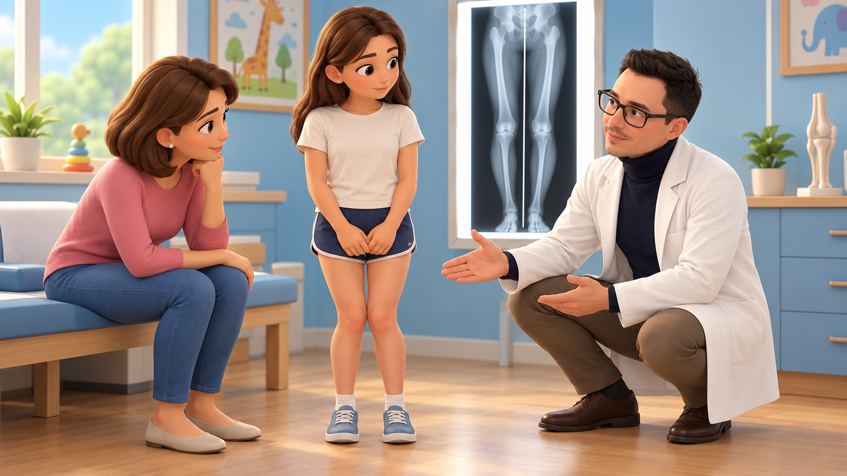
Clinical examination alone is not enough to plan treatment. Weight-bearing full-length radiographs help determine whether the deformity arises from the femur, tibia or both, and allow the mechanical axis to be measured. Remaining growth, symptoms and the adolescent’s individual circumstances also matter.
Why remaining growth changes the choice
When the growth plates are still open and enough growth remains, temporary hemiepiphysiodesis can slow one side of a growth plate while the other side continues to grow. Correction develops over the following months and requires follow-up. It is not immediate and it does not proceed at the same rate in every child.
Once the distal femoral growth plate has closed, that mechanism can no longer provide the desired correction. This does not mean that it is too late to treat the deformity. In a selected patient, it means that correction may require an osteotomy.
What a corrective osteotomy does
During a distal femoral osteotomy, the lower part of the femur is cut and realigned according to a precise plan. Internal fixation then holds the correction while the bone heals.
It is not enough to observe that the knees angle inwards. Planning must establish the site and magnitude of the deformity, joint orientation and the effect of correction on the whole limb. Multiplanar deformities and those related to skeletal dysplasia, metabolic disease or previous trauma may require a very different approach from idiopathic valgus.
A new comparison of two techniques
On 19 October 2026 the Journal of Pediatric Orthopaedics published online a randomised trial of adolescents with symptomatic idiopathic genu valgum and radiographic closure of the distal femoral physis.
The authors randomised 65 patients aged 12 to 18 years to one of two distal femoral osteotomies:
- lateral opening-wedge osteotomy;
- medial closing-wedge osteotomy.
Three patients were lost to follow-up. The available-case analysis therefore included 62 adolescents: 32 in the opening-wedge group and 30 in the closing-wedge group.
At union, mean mechanical axis deviation was 6.8 ± 6.2 mm after lateral opening wedge and 2.4 ± 5.3 mm after medial closing wedge. The difference was significant in the unadjusted analysis, but became smaller and was no longer statistically significant after covariate adjustment.
At one year there were no significant between-group differences in Pedi-IKDC functional scores, pain, satisfaction, time to union, hospital stay or return to activity. Symptomatic plate or iliotibial-band irritation occurred in 13% of the opening-wedge group and 10% of the closing-wedge group.
A few millimetres on an X-ray do not describe the whole outcome
This is the most useful part of the study. A radiographic measurement can differ between two techniques without automatically producing a benefit that the adolescent can feel.
The aim is not to chase a perfect zero on the radiograph regardless of everything else. The correction should be adequate and stable, preserve joint orientation, support functional recovery and limit complications. This trial does not establish clinically meaningful superiority of the medial closing-wedge technique and does not justify choosing an operation from a single number.
What the study cannot yet tell us
The final sample included 62 patients. That may be sufficient for a radiographic comparison, but not necessarily for uncommon complications, revisions or small functional differences. Reported functional follow-up was one year, so later differences remain uncertain, particularly in adolescents who return to high-intensity sport.
Participants had idiopathic valgus and a closed distal femoral physis. The findings should not be applied automatically to deformities associated with skeletal dysplasia, metabolic disease, previous trauma or multiplanar abnormalities.
At this stage, this commentary is based on the data available in the PubMed-indexed abstract. Critical review of the full paper remains necessary before final publication.
How the decision is made in practice
For an individual adolescent, the choice does not begin with the name of the osteotomy. It begins with the deformity.
Proper weight-bearing radiographs, measurements of the mechanical axis and joint angles, identification of the correction site and a realistic discussion about bone healing, weight-bearing, recovery and sport are required. Osteotomy geometry, fixation stability, possible changes in limb length and the team’s experience all contribute to the decision.
The new finding is reassuring without being simplistic: once growth has finished, the option to correct valgus does not disappear. The surgical tool changes. In the population studied, both techniques achieved substantial correction and favourable short- to mid-term clinical outcomes.
When assessment is useful
Orthopaedic assessment is particularly useful when valgus is marked or asymmetric, progresses over time, or is associated with pain, limping, patellar instability or activity limitation, and when it occurs in the context of skeletal disease or previous trauma.
Not every adolescent with knock knees needs an operation. The cause, severity, symptoms, skeletal maturity and the patient’s goals all matter.
Frequently asked questions
If growth is finished, can knock knees no longer be corrected?
Guided growth is no longer effective once the relevant growth plates are closed, but an osteotomy can realign the bone in selected patients. Chronological age alone does not determine the indication.
Is an osteotomy always necessary?
No. Many adolescents with mild, asymptomatic valgus do not need surgery. Deformity, symptoms, progression and functional impact must be assessed together.
Do opening-wedge and closing-wedge osteotomies give the same result?
In the new trial, both produced substantial correction and there were no significant differences in one-year functional outcomes. The study does not show that one technique is always better.
When can an adolescent return to sport after an osteotomy?
There is no universal date. Return depends on bone union, recovery of motion, strength and control, the sport involved and the treating surgeon’s instructions.
References
[1] Khatri JP, Ghosh AK, Parmar RS, et al. Lateral Opening-Wedge Versus Medial Closing-Wedge Distal Femoral Osteotomy for Adolescent Idiopathic Genu Valgum: A Randomised Controlled Trial. Journal of Pediatric Orthopaedics. Published online 19 October 2026. PubMed: 42885503 · DOI.
[2] Ranade AS, Oka GA, Belthur MV, et al. An International Consensus on Evaluation and Management of Idiopathic Genu Valgum: A Modified Delphi Survey. Journal of Pediatric Orthopaedics. 2025;45(5):274-280. PubMed: 39901614 · DOI.
[3] Artioli E, Mazzotti A, Ramacci V, et al. Indications and timing in isolated medial femoral hemiepiphysiodesis for idiopathic genu valgum: A systematic review. The Knee. 2023;40:52-62. PubMed: 36410251 · DOI.
Author: Dr Daniele Priano, Specialist in Orthopaedics and Traumatology.
Disclaimer: this article provides general information and does not replace an individual clinical assessment. The indication for correction, the technique and the postoperative pathway depend on the adolescent’s clinical and radiographic findings.
Related information
For the general clinical picture, see knock knees and bow legs in children. To understand what changes while growth is still available, see guided growth with an eight-plate and PETS or eight-plates.
The general surgical pathway is described in knock knees and bow legs: guided growth and osteotomies.