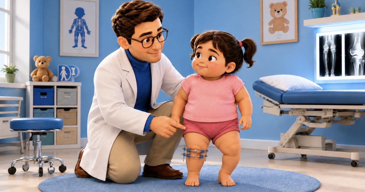
In Blount's disease, proximal tibial deformity must be considered alongside age, residual growth, weight, mechanical axis, articular component, and knee stability. Radiographic classifications remain useful, but alone are insufficient to guide treatment.
This review compiles the factors that most influence the decision between observation, bracing, guided growth, and osteotomy.
General Overview
- Blount's is not just genu varum: it is often a three-dimensional deformity with varus, procurvatum, internal tibial rotation, lateral translation, and, in advanced cases, depression of the medial tibial plateau.
- Differential diagnosis with physiological bowing remains central in early childhood: the risk is waiting too long in progressive cases.
- The metaphyseal-diaphyseal angle / Levine-Drennan angle remains a useful measurement: below 10 degrees it tends to be physiological, between 11 and 16 degrees it requires close observation, above 16 degrees it carries a high risk of progression.
- The Langenskiöld classification remains useful as a common language, but its inter-observer reliability is only moderate and does not significantly improve with MRI.
- Bracing has narrow indications: early diagnosis, young child, early stages, non-severe deformity, good family adherence.
- Guided growth remains an interesting strategy, but in Blount's, it is less predictable than in idiopathic deformities. A new study published in 2026 on juvenile and adolescent patients achieved satisfactory correction with guided growth alone in 61% of limbs; in failures, BMI was significantly higher. It is therefore a real possibility, but not a "simpler" version of osteotomy valid for everyone.
- Osteotomy remains central in progressive, severe, late-onset, multiplanar cases, or those with little residual growth.
- In advanced cases, correcting the axis is not enough if intra-articular deformity with medial tibial plateau depression is present.
- Follow-up must continue until skeletal maturity: recurrence, rebound, limb length discrepancy, and residual progression are part of the disease's history.
Recent Data on Classification and Guided Growth
The Langenskiöld classification has only moderate inter-observer agreement, even among expert evaluators. In the multicenter study by Vosoughi et al., MRI did not substantially improve its reliability. Radiographic stages must therefore be integrated with age, axis, residual growth, and clinical picture.
Guided growth is less invasive than osteotomy, but in Blount's, it utilizes a pathological physis, and results are less predictable than in idiopathic deformities. Coskun and colleagues re-evaluated 44 limbs treated with guided growth in patients with juvenile or adolescent Blount's: 61% achieved satisfactory final correction without osteotomy. In failures, the mean BMI was significantly higher. Even some severe deformities corrected, so initial severity alone does not predict the outcome.
In late-onset and adolescent forms, the deformity can involve multiple planes and segments. Evaluation must include distal femur, procurvatum/slope, rotation, limb length discrepancy, and knee stability. When residual growth, weight, three-dimensionality, or the articular component make guided growth less predictable, osteotomy offers a more controllable correction.
| Area | Message from Literature | Practical Implication |
|---|---|---|
| Classification | Langenskiöld useful but reproducibility only moderate, especially in intermediate stages. | Do not decide solely on the stage; integrate age, axis, residual growth, lateral thrust, and articular morphology. |
| Imaging | Weight-bearing radiograph remains central; MRI useful for cartilage, medial plateau, physis, and advanced cases. | Selective MRI, not routine. Useful if it changes surgical planning. |
| Guided Growth | Less predictable results in Blount's compared to idiopathic deformities. | Use in selected patients, with residual growth and non-severe deformity. |
| Osteotomy | Remains a reliable treatment for structural or progressive deformities. | Correct the coronal plane, but also rotation, procurvatum, translation, and limb length discrepancy if present. |
| Follow-up | Recurrence and rebound are frequent, especially in young, obese patients or those with significant residual growth. | Follow-up until skeletal maturity, not just until the first radiographic correction. |
Definition and Essential Pathophysiology
Blount's disease is an acquired growth disorder of the medial proximal tibia. The medial aspect of the physis slows down or loses its growth capacity compared to the lateral compartment. The consequence is a progressive varus deformity.
The most commonly used biomechanical model remains the Hueter-Volkmann principle: excessive compression reduces physeal growth. In Blount's, medial overload creates a vicious cycle: more varus, more medial load, greater growth inhibition, further worsening of the varus.
However, the mechanical component does not explain everything. Obesity, early ambulation, family predisposition, ethnic factors, and biological susceptibility of the physis contribute variably. For this reason, it is incorrect to reduce Blount's to "too much weight on the tibia."
Clinical Forms
The most practical classification distinguishes between early-onset and late-onset forms. The most commonly used cut-off is around 4 years of age.
| Form | Typical Age | Presentation | Main Therapeutic Problem |
|---|---|---|---|
| Infantile / early-onset | 1-4 years | Often bilateral, proximal tibial varus, internal torsion, possible lateral thrust; pain rare. | Early interception of progression and treatment before the deformity becomes established. |
| Juvenile | 4-10 years | Intermediate picture, often less classifiable; may represent late diagnosis of infantile form. | Often already outside the ideal window for bracing; assess residual growth and progression. |
| Adolescent / late-onset | Late childhood-adolescence | More often unilateral, associated with obesity, medial pain, limp; possible distal femoral component. | Determine if guided growth is sufficient or if osteotomy/multiplanar correction is needed. |
Diagnosis: What Should Raise Suspicion for Blount's
The greatest diagnostic difficulty is distinguishing physiological bowing from early Blount's.
Physiological bowing tends to be symmetrical, harmonious, non-progressive, and improves with growth. Blount's, on the other hand, tends to be centered on the proximal tibia, can be asymmetrical, worsens over time, and is associated with medial radiographic signs.
| Element | More Compatible with Physiological Bowing | More Suspicious for Blount's |
|---|---|---|
| Age | Especially under 2 years, with an improving trend. | Persistence after 2-3 years or worsening after the onset of walking. |
| Symmetry | Fairly symmetrical bilateral varus. | Marked asymmetry or unilateral involvement. |
| Location of Deformity | Global and harmonious limb curve. | Deformity centered on the proximal tibia. |
| Trend | Progressive clinical improvement. | Stagnation or documented worsening. |
| Dynamic Clinical Assessment | No collapse under load. | Lateral thrust during gait or stance. |
| Risk Factors | Normal weight, normal motor development. | Obesity/overweight, early ambulation, family history. |
| Radiography | Absence of metaphyseal beak, regular physis, low MDA. | Medial metaphyseal beak, physeal irregularity, elevated MDA, medial obliquity. |
Differential Diagnosis
Differential diagnosis is particularly important when the child is young, the varus is bilateral, or systemic signs are present. In these cases, it is not enough to look at the tibia: it is necessary to understand whether we are dealing with Blount's, rickets, dysplasia, or physeal sequelae.
| Differential Diagnosis | Clinical/Radiographic Clues | What to Add to Evaluation |
|---|---|---|
| Physiological Bowing | Young age, symmetrical bilaterality, harmonious curve, spontaneous improvement. | Clinical follow-up; radiograph if asymmetrical, severe, or persistent. |
| Rickets / Osteomalacia | Metaphyseal widening, diffuse alterations, short stature, pain, systemic signs. | Calcium, phosphorus, ALP, vitamin D, PTH; metabolic evaluation. |
| Skeletal/Metaphyseal Dysplasias | Disproportionate short stature, multiple deformities, family history, diffuse radiographic alterations. | Skeletal survey, genetics if indicated. |
| Traumatic or Infectious Physeal Sequelae | History of trauma, infection, surgery, progressive unilateral deformity. | Physeal evaluation, MRI/CT when appropriate. |
| Focal Bone Diseases | Metaphyseal lesions, segmental deformities, marked asymmetry. | Targeted imaging; oncological/orthopedic evaluation if atypical lesion. |
Imaging and Radiographic Measurements
Radiography remains central to diagnosis. The ideal examination is a weight-bearing full-length lower limb radiograph, with patellae facing forward. In young children, it is not always technically perfect, but one must still try to evaluate the location of the deformity, mechanical axis, and rotational component.
Elements to look for are:
- proximal tibial varus;
- medial metaphyseal beak;
- widening and irregularity of the medial physis;
- obliquity of the proximal metaphysis;
- progressive depression of the medial tibial hemi-plateau;
- lateral translation/subluxation in advanced cases;
- any associated distal femoral deformity;
- limb length discrepancy.
| Measurement | Utility | Practical Interpretation |
|---|---|---|
| Levine-Drennan / MDA | Screening in young children and distinction from physiological bowing. | <10 degrees: likely physiological; 11-16: close observation; >16: high risk of progression. |
| Mechanical Axis | Quantifies load on the medial/lateral compartment. | Fundamental for deciding treatment and correction objective. |
| MPTA | Quantifies the proximal tibial component. | Useful for planning osteotomy or guided growth. |
| LDFA | Evaluates distal femoral component. | To be measured especially in adolescents or complex deformities. |
| PPTA / Tibial Slope | Evaluates sagittal/procurvatum component. | Avoids treating only the frontal plane. |
| Limb Length Discrepancy | Evaluates length difference and planning at maturity. | Can modify choice between guided growth, osteotomy, and external fixator. |
Role of MRI
MRI is not a routine examination for every tibia vara. It becomes useful when radiography does not adequately describe the severity of the pathology or when cartilaginous/articular information changes the strategy.
Reasonable indications:
- child over 4 years old with suspected Blount's and inconclusive radiograph;
- suspected medial cartilage damage;
- suspected physeal bridge or medial bar;
- medial tibial plateau depression not well quantifiable on radiograph;
- planning of reconstructive surgery;
- discrepancy between clinical findings and radiograph.
Important limitation: MRI better visualizes cartilage, meniscus, physis, and medial plateau, but does not solve the classification problem. In the multicenter study on Langenskiöld, the addition of MRI did not substantially improve inter-observer agreement.
Classifications
Langenskiöld
The Langenskiöld classification describes six radiographic stages of the infantile form. It is still the most used system, but it should be interpreted as a descriptive language, not as a rigid therapeutic algorithm.
| Stage | Finding | Practical Implication |
|---|---|---|
| I | Medial metaphyseal irregularity. | Initial phase; possible close observation or bracing if consistent. |
| II | Medial metaphyseal beak. | Still a possible window for conservative treatment in selected young children. |
| III | Step in the metaphyseal beak. | More established deformity; often surgical indication if progressive. |
| IV | Epiphyseal involvement and initial medial depression. | The problem is no longer just extra-articular. |
| V | Double physis / advanced physeal alteration. | Risk of bar, limb length discrepancy, and recurrence; more complex surgery. |
| VI | Medial bony bar. | Advanced deformity, often requiring reconstruction; consider medial plateau, axis, and residual growth. |
Limitations of Classification
The main limitation is reproducibility. Stages II, III, and IV are the most problematic in practice. Two observers can classify the same knee differently and arrive at different indications.
For this reason, it is advisable to use three decision-making levels:
- Radiographic stage: Langenskiöld, physeal status, metaphyseal beak, medial plateau.
- Global deformity: mechanical axis, MPTA, LDFA, rotation, procurvatum, limb length discrepancy.
- Patient: age, residual growth, weight, family adherence, lateral thrust, pain, progression.
Treatment: General Principles
Treatment must be proportionate to age, residual growth, and severity of the deformity. The same radiograph can have different meanings in a 2-year-old child, a 6-year-old, and an obese adolescent near skeletal maturity.
The objectives are:
- correct the mechanical axis;
- reduce medial overload;
- correct internal tibial rotation if present;
- correct procurvatum or recurvatum if present;
- treat any associated distal femoral deformity;
- avoid or reduce final limb length discrepancy;
- prevent recurrence;
- maintain or improve knee stability and congruity.
Treatment Decision Table
| Scenario | Strategy | Notes |
|---|---|---|
| Young child, symmetrical varus, MDA <10 degrees, no suspicious radiographic signs | Observation | Clinical follow-up; radiograph if asymmetry, worsening, or persistence. |
| 2-3 years, MDA 11-16 degrees, persistent varus but not clearly progressive | Close follow-up | Gray area. Repeat clinical and imaging; do not reassure without a follow-up plan. |
| 3-4 years, Langenskiöld I-II, initial deformity, reliable family | KAFO brace | Narrow window. If no improvement, switch to surgical strategy without delay. |
| Age >3-4 years, progression, Langenskiöld III or lateral thrust | Proximal tibial osteotomy | Correct axis, rotation, and sagittal component if present. |
| Adolescent with mild-moderate varus, sufficient residual growth, predominantly coronal deformity | Selective guided growth | Requires serial check-ups; attention to failure, rebound, and implant breakage. |
| Obese adolescent, severe varus, multiplanar deformity or little residual growth | Osteotomy, often with gradual correction | Consider circular/hexapod fixator if complex correction or associated with limb length discrepancy. |
| Langenskiöld V-VI, medial plateau depression, lateral thrust or incongruity | Reconstructive surgery | Possible medial plateau elevation + realignment + growth/limb length discrepancy management. |
Observation
Observation is indicated only if the picture is compatible with physiological bowing or with very doubtful but non-progressive Blount's.
Criteria favorable for observation:
- young age;
- symmetrical varus;
- absence of lateral thrust;
- MDA less than 10 degrees;
- no significant medial metaphyseal beak;
- improving clinical course;
- no significant risk factors.
In the gray area, observation must be active: clinical follow-up and, when indicated, repeat radiographs. The point is not to avoid radiographs at all costs; the point is not to miss progression.
Bracing
Bracing is indicated primarily in the early infantile form. The ideal candidate is a child diagnosed early, with Langenskiöld I-II, mild-moderate deformity, absence of significant lateral thrust, and good family compliance.
The brace is generally a KAFO applying a corrective valgus force. Treatment requires time and adherence. Literature emphasizes that success is greater if started before 3 years of age and in non-obese children. If the brace does not lead to improvement, osteotomy should not be delayed too long.
| Brace: Favorable Indications | Brace: Unfavorable Signs |
|---|---|
| Age <3 years | Age >3-4 years |
| Langenskiöld I-II | Langenskiöld III or higher |
| Mild-moderate deformity | Severe or progressive varus |
| No significant lateral thrust | Obvious lateral thrust |
| Cooperative family | Poor predictable adherence |
| Non-obese child | Significant obesity |
Guided Growth
Guided growth is conceptually attractive because it is less invasive than osteotomy. In Blount's, however, it should not be trivialized.
In idiopathic genu varum, a healthy physis that continues to grow is exploited. In Blount's, on the other hand, the medial physis is pathological or otherwise biologically unfavorable. For this reason, correction can be slow, incomplete, or recurrent.
More reasonable indications:
- late-onset Blount's or adolescent with residual growth;
- mild-moderate deformity;
- predominantly coronal varus;
- absence of severe medial plateau depression;
- absence of significant lateral thrust;
- at least 2 years of residual growth, preferably more;
- possibility of close follow-up.
Cases where caution is needed:
- obese adolescent with severe varus;
- multiplanar deformity;
- little residual growth;
- severe involvement of the medial physis;
- breakage or high mechanical risk of the implant;
- family expectation of "simple and definitive" correction.
| Advantages of Guided Growth | Limitations of Guided Growth in Blount's |
|---|---|
| Less invasiveness | Correction dependent on residual growth |
| Lower morbidity compared to osteotomy | Unpredictable response of the pathological medial physis |
| Possible gradual correction without osteotomy | Possible incomplete correction |
| Implant removal upon correction achievement | Risk of rebound/recurrence |
| Useful in mild-moderate deformities with residual growth | Risk of screw/implant breakage in heavy patients or severe deformities |
Proximal Tibial Osteotomy
Osteotomy remains the cornerstone treatment for structural, progressive, or severe deformities. It is indicated when the problem cannot be reliably solved with bracing or growth modulation.
Typical indications:
- age over 3-4 years with progression;
- Langenskiöld III or higher;
- brace failure;
- significant varus;
- lateral thrust;
- multiplanar deformity;
- little residual growth;
- severe adolescent Blount's;
- guided growth not indicated or insufficient.
Planning must consider:
- level of deformity;
- mechanical axis;
- MPTA and LDFA;
- tibial rotation;
- procurvatum/recurvatum;
- limb length discrepancy;
- physeal status;
- medial plateau status;
- neurovascular risk.
In the surgically treated infantile form, overcorrection into valgus is often discussed to reduce the risk of recurrence. This should not be interpreted as a fixed number, but as a principle: the load must be shifted out of the pathological medial compartment.
Acute Correction and Gradual Correction
Acute correction is useful in deformities that are not excessively complex, with the possibility of achieving adequate axis and rotation without excessive neurovascular risk.
Gradual correction with a circular or hexapod fixator is more demanding but becomes very useful when the deformity is severe, multiplanar, or associated with limb length discrepancy.
| Parameter | Acute Correction | Gradual Correction |
|---|---|---|
| Deformity | Mild-moderate, relatively simple. | Severe, multiplanar, recurrent, or complex. |
| Rotation/Procurvatum | Correctable if well planned. | More controllable progressively. |
| Limb Length Discrepancy | Does not correct large differences well. | Can be associated with lengthening. |
| Neurological Risk | Higher with large acute correction. | Reduced by gradualness, but not zero. |
| Family Management | Simpler after surgery. | More complex: dressings, check-ups, adjustments. |
| Typical Complications | Loss of correction, common peroneal nerve palsy, compartment syndrome, problems with bone healing. | Pin-site infection, pain, stiffness, compliance, treatment duration. |
Articular Deformity and Medial Tibial Plateau Elevation
In advanced forms, especially Langenskiöld V-VI, the problem is no longer solely extra-articular. If the medial tibial plateau is depressed, the medial femur continues to articulate on an incongruent surface. In these cases, realignment osteotomy alone may not resolve knee stability.
Signs that should suggest an articular component:
- obvious medial depression;
- persistent lateral thrust;
- articular incongruity;
- advanced stages;
- pain and instability;
- subluxation/lateral translation.
In selected cases, medial tibial plateau elevation should be considered, associated with realignment, management of residual growth when indicated, and treatment of limb length discrepancy.
Complications and Failures
The management of Blount's must include, from the outset, a realistic discussion of complications and the need for staged treatments.
| Strategy | Possible Problems | Prevention/Management |
|---|---|---|
| Observation | Diagnostic delay, unrecognized progression. | Scheduled follow-up, radiographic measurements, serial comparison. |
| Bracing | Poor compliance, ineffectiveness in late cases, loss of surgical window. | Strict indication and early re-evaluation of effectiveness. |
| Guided Growth | Incomplete correction, rebound, screw/implant breakage, overcorrection. | Patient selection, serial radiographic follow-up, timely removal. |
| Acute Osteotomy | Common peroneal nerve palsy, compartment syndrome, loss of correction, recurrence. | Accurate planning, neurovascular protection, fasciotomy when appropriate. |
| External Fixator | Pin-site infection, pain, stiffness, family intolerance. | Family education, frequent check-ups, physiotherapy, pin-site management. |
| Medial Plateau Elevation | Stiffness, residual incongruity, surgical complexity. | Selective indication and three-dimensional planning. |
Follow-up
Follow-up is part of the treatment. It is not enough to correct the axis once.
Elements to monitor:
- mechanical axis;
- MDA/MPTA/LDFA according to age;
- tibial rotation;
- lateral thrust;
- pain;
- limb length discrepancy;
- implant status;
- rebound after guided growth removal;
- varus recurrence;
- skeletal maturity.
A possible practical schedule:
| Phase | Follow-up | Objective |
|---|---|---|
| Initial Observation | 3-6 months depending on risk. | Understand if varus improves, remains stable, or progresses. |
| Bracing | Close follow-up, often 3-4 months. | Verify compliance and correction; do not waste time if ineffective. |
| Guided Growth | Serial X-rays every 3-4 months approximately. | Monitor correction, avoid overcorrection, identify breakages. |
| Post-Osteotomy | Until consolidation, then growth follow-up. | Verify axis, consolidation, recurrence, limb length discrepancy. |
| Until Maturity | Annually or according to risk. | Detect rebound, recurrence, and final limb length discrepancy. |
Final Summary Table
| Clinical Question | Practical Answer |
|---|---|
| Is it physiological? | Only if symmetrical, harmonious, non-progressive, low MDA, and without medial signs. |
| When should I be concerned? | Asymmetry, worsening, proximal tibia, MDA >16 degrees, lateral thrust, obesity, medial radiographic signs. |
| When to brace? | Young child, preferably <3 years, Langenskiöld I-II, initial deformity, and reliable family. |
| When guided growth? | Mild-moderate deformity, residual growth, predominantly coronal picture, no severe medial depression. |
| When osteotomy? | Progression, late age, stage III or higher, lateral thrust, severe or multiplanar deformity. |
| When fixator? | Severe deformity, recurrence, obese adolescent, multiplanar correction, or need for lengthening. |
| When to consider the medial plateau? | Langenskiöld V-VI, medial depression, incongruity, persistent lateral thrust. |
Conclusions
Modern management of Blount's disease cannot be reduced to "radiographic stage equals treatment." Classification helps, but does not decide alone.
Correct treatment arises from the integration of:
- age;
- residual growth;
- progression;
- MDA and mechanical axis;
- severity of deformity;
- three-dimensional component;
- status of the medial physis;
- status of the tibial plateau;
- body weight;
- family compliance;
- risk of recurrence.
Recent literature makes the automatic use of Langenskiöld more cautious and the use of guided growth more selective. At the same time, it confirms that bracing, hemiepiphysiodesis, acute osteotomy, gradual correction, and articular surgery all have a role, but in different windows of the disease.
The final message is simple: in Blount's, timing weighs as much as technique. A "less invasive" treatment indicated late can be worse than a more demanding surgery indicated correctly.
Bibliography
- Blount WP. Tibia vara: osteochondrosis deformans tibiae. J Bone Joint Surg. 1937.
- Langenskiöld A. Aspects of the pathology of tibia vara. Ann Chir Gynaecol Fenn. 1955.
- Davids JR, Blackhurst DW, Allen BL. Radiographic evaluation of bowed legs in children. J Pediatr Orthop. 2001;21(2):257-263.
- Sabharwal S, Zhao C, McClemens E. Correlation of body mass index and radiographic deformities in children with Blount disease. J Bone Joint Surg Am. 2007;89(6):1275-1283.
- Sabharwal S. Blount disease. J Bone Joint Surg Am. 2009;91(7):1758-1776.
- Birch JG. Blount disease. J Am Acad Orthop Surg. 2013;21(7):408-418.
- Sabharwal S. Blount disease: an update. Orthop Clin North Am. 2015;46(1):37-47.
- Sabharwal S, Sabharwal S. Treatment of Infantile Blount Disease: An Update. J Pediatr Orthop. 2017;37 Suppl 2:S26-S31.
- Janoyer M. Blount disease. Orthop Traumatol Surg Res. 2019;105(1S):S111-S121.
- Park BK, Park KB, Kwak YH, Jin S, Kim HW, Park H. A comparative evaluation of tibial metaphyseal-diaphyseal angle changes between physiologic bowing and Blount disease. Medicine (Baltimore). 2019;98(17):e15349.
- Erkus S, Turgut A, Kalenderer O. Langenskiöld Classification for Blount Disease: Is It Reliable? Indian J Orthop. 2019;53(5):662-664.
- Vosoughi F, Nabian MH, Simon AL, et al. Langenskiöld classification of tibia vara: a multicenter study on interrater reliability. J Pediatr Orthop B. 2022;31(2):114-119.
- Schroerlucke S, Bertrand S, Clapp J, Bundy J, Gregg FO. Failure of Orthofix eight-Plate for the treatment of Blount disease. J Pediatr Orthop. 2009;29(1):57-60.
- Stitgen A, Garrels K, Kobayashi H, Vanderby R, McCarthy JJ, Noonan KJ. Biomechanical comparison between 2 guided-growth constructs. J Pediatr Orthop. 2012;32(2):206-209.
- Burghardt RD, Herzenberg JE, Strahl A, Bernius P, Kazim MA. Treatment failures and complications in patients with Blount disease treated with temporary hemiepiphysiodesis: a critical systematic literature review. J Pediatr Orthop B. 2018.
- Fan B, Zhao C, Sabharwal S. Risk factors for failure of temporary hemiepiphysiodesis in Blount disease: a systematic review. J Pediatr Orthop B. 2020.
- Feldman DS, Madan SS, Ruchelsman DE, Sala DA, Lehman WB. Accuracy of correction of tibia vara: acute versus gradual correction. J Pediatr Orthop. 2006;26(6):794-798.
- Griswold B, Gilbert S, Khoury J. Opening Wedge Osteotomy for the Correction of Adolescent Tibia Vara. Iowa Orthop J. 2018;38:141-146.
- Dakshina Murthy TSS, Taqi M, De Leucio A. Blount Disease. StatPearls. Updated 2024.
Bibliographic Update 2026
Coskun E, Bess LE, Bielski RJ. The Outcomes of Guided Growth System in Juvenile and Adolescent Patients With Blount Disease. J Pediatr Orthop. 2026;46(3):166-171. doi:10.1097/BPO.0000000000003149. PMID: 41178588.
