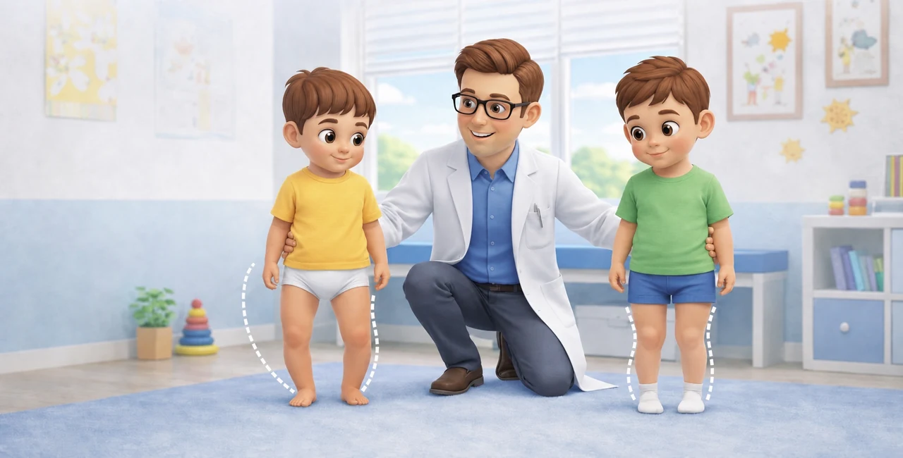
In the first years of life, leg alignment changes significantly. A phase of bow-leggedness (varus) is common in young children; subsequently, the axis tends towards neutral and then to a physiological knock-knee (valgus), more evident around 3–4 years of age, which progressively reduces towards adult values. [1–3]
For this reason, an 18-month-old child with bow legs and a 4-year-old with knock knees can both be within a normal growth phase.
When the condition is likely physiological
Symmetry is one of the most useful elements. Bilateral, harmonious bow-leggedness or knock-knees, without pain and consistent with age, is generally reassuring. The progression over time also matters: physiological deformities tend to follow a predictable trajectory and do not progressively worsen. [1–3]
In typical cases, orthotics, corrective shoes, or braces are not needed to "straighten" the legs faster. There is no evidence that these aids change the natural history of physiological alignment. [1,3]
When it's worth investigating further
Marked asymmetry, worsening, pain, limping, short stature, an uneven growth pattern, or persistence of a significant deformity beyond the expected age warrant a more in-depth evaluation. [1,2,4]
In a young child with genu varum (bow legs), one of the diagnoses to distinguish is Blount's disease. In these cases, the course and, when indicated, a weight-bearing X-ray help differentiate the physiological variant from a pathological deformity of the proximal tibia. [1]
The role of vitamin D
A small Japanese study from 2025 evaluated vitamin D supplementation in 90 children between 10 and 30 months with genu varum and observed greater radiographic improvement in the treated group. [4]
This is interesting but preliminary data. It does not justify automatic testing or supplementation in every child with bow legs. Metabolic tests become more sensible when the condition is marked, progressive, or associated with short stature, pain, metaphyseal alterations, or other elements that suggest rickets or a metabolic disorder.
What I look for during the visit
Age, symmetry, location of the deformity, stature, progression over time, and presence of pain or functional difficulties are more informative than just the aesthetic impression.
Many children between 0 and 6 years old do not need any treatment. Orthopedic check-ups primarily serve to recognize the few cases where the deformity does not follow the normal course of growth.
Disclaimer
This content is for informational purposes only and does not replace an individual clinical evaluation.
References
[1] Coppa V, Marinelli M, Procaccini R, Falcioni D, Farinelli L, Gigante A. Coronal plane deformity around the knee in the skeletally immature population: A review of principles of evaluation and treatment. World J Orthop. 2022;13(5):427-443. doi:10.5312/wjo.v13.i5.427. PMID: 35633744.
PubMed: https://pubmed.ncbi.nlm.nih.gov/35633744/
[2] Patel M, Nelson R. Genu Valgum. StatPearls [Internet]. Updated 2025 Jan. PMID: 32644670.
PubMed: https://pubmed.ncbi.nlm.nih.gov/32644670/
[3] Weiner DS. The natural history of “bow legs” and “knock knees” in childhood. Orthopedics. 1981;4(2):156-160. doi:10.3928/0147-7447-19810201-08. PMID: 24823174.
PubMed: https://pubmed.ncbi.nlm.nih.gov/24823174/
[4] Sakamoto Y, Kamegaya M, Saisu T, Tomaru Y, Tokita A, Kim SG, Ishijima M. Vitamin D supplementation improves genu varum in toddlers: two-center pilot study. J Bone Miner Metab. 2025;43(3):265-273. doi:10.1007/s00774-025-01583-1. PMID: 39918569.
